
Veterinary Neurology and Neurosurgery
Our team is dedicated to diagnosing and treating a wide range of neurological disorders in dogs and cats, working closely with our Physical Therapy & Rehabilitation team to achieve the best possible outcomes.
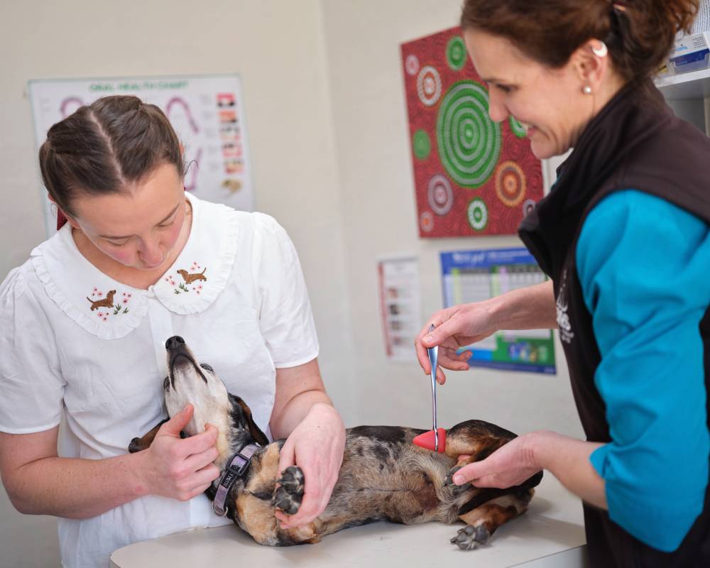
Neurosurgery at BVRS
At BVRS, we offer a range of spinal surgeries, most commonly spinal decompression for intervertebral disc disease (IVDD).
IVDD – Intervertebral Disc Disease
What is IVDD?
Intervertebral disc disease (IVDD) is the most frequently encountered spinal disorder in dogs and can also occur in cats. The intervertebral discs are fibrocartilaginous structures located between most vertebrae, with the exception of the joint between C1 and C2. They provide support and flexibility to the spine while also helping to absorb mechanical forces. When these discs degenerate, their normal structure and function are progressively altered, making them more prone to disc herniation and spinal cord compression.
Causes
IVDD develops as a consequence of degenerative changes within the intervertebral discs. In some dogs, particularly those classified as chondrodystrophic, these changes can begin at a relatively young age. The centre of a healthy disc has a well-hydrated, jelly-like consistency; with degeneration and dehydration, it becomes more like a gritty paste. This change alters its function, decreasing its ability to be flexible and absorb shock, and makes it more likely to result in disc bulging or herniation.
Chondrodystrophic breeds have characteristically shortened limbs in proportion to their body length. Dachshunds, Beagles and many Terrier breeds are common examples. These dogs are predisposed to developing early degenerative changes within their intervertebral discs, increasing the likelihood of subsequent disc-related disease.
Clinical signs
Dogs and cats with IVDD may initially develop pain localised to the spine, either in the neck or back. Neurological dysfunction can subsequently develop, ranging from mild weakness through to complete paralysis. In some patients, sudden paralysis may be the first obvious indication of a spinal problem.
Because the spinal cord carries nerve pathways between the brain and the limbs and organs, the location of the disc lesion determines which parts of the body are affected. For example, disease affecting the lower back may result in hindlimb weakness or paralysis and may interfere with normal bladder function. A disc problem within the cervical spine can affect all four limbs and may result in weakness or paralysis.
Pain is commonly one of the earliest clinical signs. Affected animals may adopt an abnormal or hunched posture, tremble, pant, become reluctant to move, or have difficulty jumping onto furniture or climbing stairs. As the condition progresses, animals may develop an unsteady gait, reduced limb coordination, weakness, ataxia or paralysis. Severe spinal cord injury can also result in impaired bladder emptying and loss of sensation in the limbs.
Spinal cord disease should be considered an emergency. Veterinary assessment by a Specialist Surgeon should be sought promptly following the development of neurological or spinal pain signs, as delays in treatment may negatively affect the prognosis.
Diagnosis
IVDD may be suspected from the clinical examination and the patient’s breed and history, but imaging is required to establish the diagnosis and determine the exact location and severity of the lesion. Standard spinal radiographs can demonstrate changes associated with disc degeneration, but they generally do not provide sufficient information about the spinal cord or the precise location of compression to allow appropriate surgical planning.
Advanced diagnostic imaging is therefore required for definitive assessment. Depending on the individual patient, myelography, computed tomography (CT) or magnetic resonance imaging (MRI) may be recommended. The most appropriate imaging technique will be selected by the surgeon according to the clinical presentation and suspected location of the lesion.
Advanced imaging provides considerably more information than routine radiographs and allows the BVRS team to identify the site of spinal cord compression and formulate an appropriate treatment plan. Because the patient must remain completely still during these procedures, deep sedation or general anaesthesia is required. Without advanced imaging, an accurate diagnosis and appropriate treatment plan cannot reliably be established.
Differential diagnoses
A number of other diseases can produce clinical signs that resemble IVDD. These include inflammatory conditions such as meningitis, infection involving the vertebrae or intervertebral discs, spinal tumours or other cancers, vascular events involving blood clots (spinal stroke) or cartilage clots (FCE), traumatic spinal injury and paralysis caused by tick envenomation.
Because these conditions can have very similar clinical presentations, diagnostic imaging such as myelography, CT or MRI is often necessary to distinguish between them and identify the underlying cause.
Treatment
Conservative or Non-surgical
Non-surgical treatment may be appropriate for patients whose primary clinical sign is pain or whose neurological deficits are very mild. Animals with more substantial neurological impairment are generally less likely to achieve a satisfactory recovery with conservative treatment alone. Loss of pain sensation indicates a severe spinal cord injury and represents a surgical emergency; these patients have an extremely poor likelihood of successful recovery with conservative treatment.
A further limitation of conservative management is the increased risk of recurrent clinical signs, ongoing neurological abnormalities or deterioration.
The main benefit of conservative treatment is that it avoids surgery and is generally less costly. Strict restriction of activity is essential and typically involves cage or crate confinement for at least eight weeks with regular physical therapy visits. Limiting movement reduces the risk of further disc material extrusion and allows the damaged disc and spinal cord to undergo natural healing. Appropriate analgesia is an important component of treatment, and patients should undergo regular veterinary reassessment to monitor their neurological status and recovery.
Surgical Treatment
Surgical decompression is an important treatment option for dogs and cats with significant neurological deficits caused by IVDD. Early surgical treatment is associated with a greater likelihood of achieving a rapid and complete return of neurological function.
Although some patients can recover with conservative treatment, surgery may provide a more favourable outcome in appropriately selected cases. Potential benefits include a greater likelihood of improvement, faster neurological recovery, improved pain control and a reduced risk of recurrence at that location. Patients that have lost deep pain sensation require immediate surgical attention and have a poor prognosis for meaningful improvement without prompt decompression.
What does the surgery involve?
The primary goal of surgery is to relieve pressure on the spinal cord by removing disc material that has entered the vertebral canal. These procedures require specialised equipment, technical expertise and appropriate surgical training.
The surgical approach selected depends largely on the location of the disc extrusion. For cervical disc disease, a ventral approach is commonly used. This procedure, often referred to as a ventral slot, involves creating an opening through the vertebral bodies to access and remove the affected disc material.
For lesions affecting the thoracolumbar spine, a hemilaminectomy is frequently performed. This approach accesses the vertebral canal from the side of the vertebra, directly over the affected disc space.
At the lumbosacral junction, a dorsal laminectomy may be indicated. This involves removing the top of the vertebral canal to allow access to the nerves and affected lumbosacral disc.
Other surgical techniques may also be appropriate depending on the location and characteristics of the disc disease. The BVRS team will discuss the procedure considered most suitable for your individual pet.
Rehabilitation
Rehabilitation Overview
After surgery, your pet will be assessed by our Rehabilitation team. An individual program will be developed based on their neurological function, strength, mobility and specific recovery needs.
Rehabilitation is an important component of recovery following spinal cord disease. Prolonged inactivity or recumbency can contribute to reduced joint mobility, stiffness, muscle loss and contracture. Appropriate physiotherapy helps maintain mobility and muscle function while supporting the return of normal movement.
How long before my paralysed pet will walk again?
Recovery following severe spinal cord injury is highly variable. Some animals begin to regain function relatively quickly, while others may require several weeks or longer before meaningful improvement becomes apparent.
The rate and extent of recovery depend on the severity and location of the spinal cord injury and the neurological function present at the time of treatment. It is important to allow sufficient time for recovery, as neurological improvement can continue over an extended period. Unfortunately, a small proportion of severely affected patients may not regain the ability to walk.
Recurrence
Recurrence at the original surgical site is uncommon following successful decompression. However, animals with degenerative disc disease remain susceptible to problems involving other intervertebral discs.
The likelihood of developing disease at another spinal level varies between breeds and individual patients and is most common in Dachshunds.
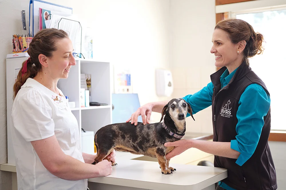
Services may include the investigation and treatment of:
- Intervertebral disc disease (IVDD)
- Degenerative lumbosacral stenosis
- Cervical spondylomyelopathy (wobbler syndrome)
- Subarachnoid diverticulum
- Spinal fractures

Contact BVRS About Neurology and Neurosurgery
If your pet has been referred for Neurology and Neurosurgery, or if you are a veterinarian seeking specialist surgical support, please contact our team.